C6, 9L, and F98 are the three of the most used rat glioma models in research. In their recent review article, Sahu et al. describe the origin, genomics, and use of these three models in research1. To this day, gliomas remain incredibly hard to treat and patients commonly have very low survival rates. Orthotopic rat brain cancer models are central to generating better clinical interventions. Leveraging our expertise in genetically engineered cell-lines, rat surgeries, and in vivo bioluminescent imaging capabilities, Hera BioLabs is expanding our orthotopic brain offerings in rats.
Some of the advantages of rat versus mouse brain tumor models are as follows:
- A larger brain allows more precise stereotactic implantation and a longer time until tumor endpoint
- The larger tumor size creates better in vivo imaging
- Therapeutic agents can be given intracerebrally (i.c.) with greater ease
- More literature and research exist on rat brain tumors compared to their mouse counterparts
Hera BioLabs’s SRG Rat™
We offer another significant advantage – a highly immunocompromised rat model, the SRG. Lack of a complete immune system can counteract the inability to generate orthotopic data as two of these most widely used glioma models, C6 and 9L are highly immunogenic in many rat strains. If an intact immune system is not required, the SRG rat facilitates studies using immunogenic cell lines.
The C6 cell line is the most used rodent brain tumor model and expresses genes resembling those reported in human gliomas. Several of these genes are responsible for stimulation of the Ras pathway. C6 is widely used to evaluate therapeutic efficacy in vitro, yet it is immunogenic even in its host Wistar rat and currently has no host it can be successfully propagated in.
9L is the second most commonly used rat brain tumor model. 9L is a gliosarcoma derived from Fischer rats and develops rapidly growing tumors when implanted i.c. This cell line is primarily used to study drug transport in the brain and tumor blood barriers, drug resistance, modeling brainstem tumors, and MRI, PET, and PK studies. Similar to C6, 9L can provoke a strong antitumorigenic immune response.
F98 glioma is the third most common rat brain tumor model. Genes that are overexpressed in F98 include Ras, PDGFβ, cyclin D1/D2, EGFR, and Rb. This model simulates human GBMs closer than other cell lines due to its high invasiveness and weak immunogenicity, making it an ideal candidate for studying efficacy of therapeutic agents. F98 also has several gene-modified versions including F98-EGFR, F98npEGFRvlll, and a luciferase expressing line which allows for in-vivo imaging.

Table 1: Comparison of C6, 9L, and F98 significant molecular markers.

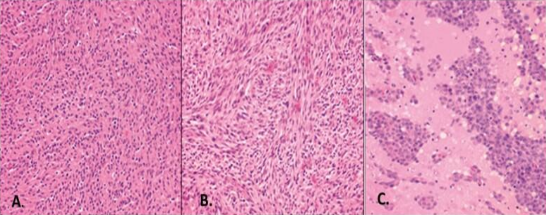
Figure 1: Histopathological view of the C6, 9L, and F98 rat brain tumors. A. C6 glioma is composed of pleomorphic cells ranging from round to oblong nuclei with a mild herring pattern of growth and focal invasion of contiguous normal brain. B. 9L gliosarcoma is composed of spindle-shaped cells with a sarcomatoid appearance. A whorled pattern of growth is seen, with little invasion of contiguous normal brain. C. F98 glioma is composed of a mixed population of spindle-shaped cells with fusiform nuclei, a frequent whorled pattern of growth is seen with a smaller subpopulation of polygonal cells with round to oval nuclei. Extensive invasion of the contiguous brain occurs with islands of tumor cells and a central area of necrosis.
Work With Hera BioLabs
Reach out to us to learn more about using our SRG Rat for your research.
References
- Sahu, U., Barth, R. F., Otani, Y., McCormack, R. & Kaur, B. Rat and Mouse Brain Tumor Models for Experimental Neuro-Oncology Research. J Neuropathol Exp Neurol 81, 312-329, doi:10.1093/jnen/nlac021 (2022).
