Organ Microenvironment In Cancer Growth And The Importance Of In Vivo Research
Over 130 years ago, Stephen Paget analyzed autopsy data from 735 women with breast cancer and observed non-random tumor metastases to specific tissues. Thus, Paget hypothesized the “seed and soil” model of tumor metastasis wherein tumor cells preferentially invade and grow in the microenvironment of specific organs. In the following decades, thousands of studies have supported the essential role of the tumor microenvironment in the progression of both primary tumors and metastases.
Over the past 5 decades, several immunodeficient rodent models have been used for propagating human cancer xenografts. At the culmination of this research is Hera BioLabs’ Sprague Dawley Rag2 -/-, Il2rg -/- rat, the SRGTM rat (OncoRat®)1. In rodent models, subcutaneous tumor inoculations are often appropriate for testing anticancer drug efficacy. However, numerous studies have shown that rodent models of orthotopic tumor inoculations more closely recapitulate the natural history of cancer in humans, including higher rates of metastasis2. Thus, in vivo assessment of primary tumor growth and metastasis in rodents is essential for understanding the mechanisms of tumor progression and dissemination.
Bioluminescent Cell Line Imaging
Leveraging our expertise in cell line engineering using the piggyBac transposon system, Hera BioLabs is developing a panel of popular cancer cell lines with stable Firefly Luciferase (FLuc) expression. To track our FLuc-tagged tumor lines in vivo, we now offer in vivo imaging using the AMI HT platform from Spectral Instruments imaging.

The AMI HT imager has numerous convenient features, including:
- Bioluminescent or fluorescent imaging
- Cutting edge patented LED based illumination
- 10 Wavelengths/10 Filters
- Ultra-cold camera for maximum sensitivity
- Absolute Calibration ensures quantifiable data
- Aura Imaging Software–100% license free
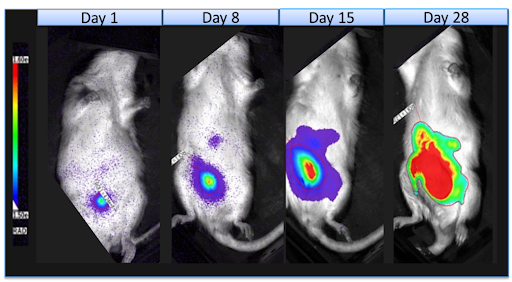
The SRGTM rat lacks B, T, and NK cells, and supports robust growth of multiple human cancer cell lines. In an initial proof-of-principle study, Hera established orthotopic OV81.2-luc (a human serous ovarian carcinoma cell line established from a patient ascites tumor sample and engineered to have stable luciferase expression) tumors in female SRG rats by intraperitoneal injection. Tumor growth was tracked with weekly in vivo luciferase imaging using the AMI HT system. As seen in Figure 1, luciferase signal was detectable 1 day after initial injection, and tumor growth was observed at 8, 15, and 28 days after injection. At necropsy, we found no evidence of metastases outside of the peritoneal cavity, but we observed tumors on abdominal organs including ovary, mesentery, intestines, kidneys, liver, and abdominal wall.

Figure 2: in-vivio-luminescent-imaging-in-the-SRG-OncoRat
Five million OV81.2-luc ovarian cancer cells were injected i.p. into SRG rats (N=3), and imaging was used to track tumor growth.
These data confirm the feasibility and utility of studies that combine bioluminescent cancer cell lines with in vivo imaging in the SRGTM OncoRat®. Importantly, the larger size of the rats (versus mice) increases the ease of surgical procedures necessary for orthotopic implantation, allowing for models that closely recapitulate human cancers and their microenvironment. The SRGTM OncoRat® has other advantages over mice, including the ability to combine drug efficacy, tumor growth kinetics, and PK/PD studies. Larger tumor volumes allow for serial biopsies for detailed molecular characterization of tumors throughout the treatment regimen.
Thus, in vivo bioluminescent imaging in the SRGTM OncoRat® provides an ideal platform for de-risking your pipeline and developing life-saving pharmacotherapies.
If your preclinical studies could use a boost, or you would like to see more data on the SRG Rat, contact Hera BioLabs for more information here.
References
- Noto, F. K. et al. The SRG rat, a Sprague-Dawley Rag2/Il2rg double-knockout validated for human tumor oncology studies. PLoS One 15, e0240169, doi:10.1371/journal.pone.0240169 (2020).
- Ruggeri, B. A., Camp, F. & Miknyoczki, S. Animal models of disease: pre-clinical animal models of cancer and their applications and utility in drug discovery. Biochem Pharmacol 87, 150-161, doi:10.1016/j.bcp.2013.06.020 (2014).
